|

HEALTH EFFECTS: Fluoride & the Reproductive System
Key Findings - Fluoride & the Reproductive System:
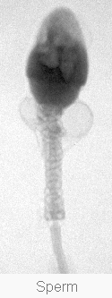 High doses of fluoride have repeatedly been found to interfere with the reproductive system of animals . Commonly observed effects in fluoride-exposed animals include: oxidative stress, damaged sperm, reduced sperm count, and reduced fertility.
According to the authors of a recent study in the journal Reproductive Toxicology :
"We conclude that fluoride treatment is associated with testicular disorders, which may be due to induction of oxidative stress in reproductive organs along with possible adverse effects of fluoride on pituitary testicular axis.The detailed mechanism of fluoride treatment on the male reproductive system has not been elucidated and will be the subject of future experiments " (Ghosh et al 2002).
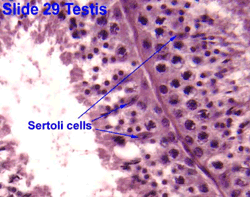
Research on possible reproductive effects in humans is limited. Some recent research , however, indicates that fluoride exposure (at lower doses than given to animals) can cause toxic effects to human Sertoli cells and gonadotrophs, reduction in circulating testosterone, and reductions in total fertility rate. The dose at which fluoride can begin to cause these effects is not yet known.
Need for Research - Fluoride & the Reproductive System: ( back to top )
the relationship between fertility and fluoride requires additional study.
SOURCE: National Research Council. (2006). Fluoride in Drinking Water: A Scientific Review of EPA's Standards . National Academies Press, Washington D.C. p161.
"The effects of fluoride on the reproductive system merit further investigation in animal and human studies."
SOURCE: Department of Health & Human Services. (U.S. DHHS) (1991). Review of Fluoride: Benefits a nd Risks . Department of Health and Human Services, USA. p. 88-89.
Human Studies - Fluoride & the Reproductive System: ( back to top )
 A few human studies suggested that high concentrations of fluoride exposure might be associated with alterations in reproductive hormones, effects on fertility, and developmental outcomes, but design limitations make those studies insufficient for risk evaluation.
SOURCE: National Research Council. (2006). Fluoride in Drinking Water: A Scientific Review of EPA's Standards . National Academies Press, Washington D.C. p 6.
"Fluoride-induced reproductive effects have been reported in experimental models and in humans. However, these effects were found in heavily exposed scenarios. Therefore, in this work our objective was to study reproductive parameters in a population exposed to fluoride at doses of 3-27 mg/day (high-fluoride-exposed group-HFEG). Urinary fluoride levels, semen parameters, and reproductive hormones in serum (LH, FSH, estradiol, prolactin, inhibin-B, free and total testosterone) were measured. Results were compared with a group of individuals exposed to fluoride at lower doses: 2-13 mg/day (low-fluoride-exposed group-LFEG). A significant increase in FSH (P<0.05) and a reduction of inhibin-B, free testosterone, and prolactin in serum (P<0.05) were noticed in the HFEG. When HFEG was compared to LFEG, a decreased sensitivity was found in the FSH response to inhibin-B (P<0.05). A significant negative partial correlation was observed between urinary fluoride and serum levels of inhibin-B (r=-0.333, P=0.028) in LFEG. Furthermore, a significant partial correlation was observed between a chronic exposure index for fluoride and the serum concentrations of inhibin-B (r=-0.163, P=0.037) in HFEG. No abnormalities were found in the semen parameters studied in the present work, neither in the HFEG, nor in the LFEG. The results obtained indicate that a fluoride exposure of 3-27 mg/day induces a subclinical reproductive effect that can be explained by a fluoride-induced toxic effect in both Sertoli cells and gonadotrophs."
SOURCE: Ortiz-Perez D, et al. (2003). Fluoride-induced disruption of reproductive hormones in men. Environmental Research 93:20-30.
"The first step in assessing a health risk by a substance to humans is the identification of its harmful effects on animals . A health risk to humans is assessed using results from human epidemiological studies in conjunction with results from animal studies. The Newburgh-Kingston Study (Schlesinger et al, 1956) showed an earlier age of first menarche in girls living in the fluoridated Newburgh than in unfluoridated Kingston. The current animal study indicates that fluoride is associated with an earlier onset of puberty in female gerbils. Furthermore, more research was recommended on the effects of fluoride on animal and human reproduction (USPHS, 1991). This project has contributed new knowledge in this area."
SOURCE: Luke J. (1997). The Effect of Fluoride on the Physiology of the Pineal Gland . Ph.D. Thesis. University of Surrey, Guildford. p. 177.
SOURCE: Susheela AK, Jethanandani P. (1996). Circulating testosterone levels in skeletal fluorosis patients. Journal of Toxicology and Clinical Toxicology 34(2):183-9.
"A review of fluoride toxicity showed decreased fertility in most animal species studied. The current study was to see whether fluoride would also affect human birth rates. A U.S. database of drinking water systems was used to identify index counties with water systems reporting fluoride levels of at least 3 ppm. These and adjacent counties were grouped in 30 regions spread over 9 states... Most regions showed an association of decreasing TFR [Total Fertility Rate] with increasing fluoride levels. Meta-analysis of the region-specific results confirmed that the combined result was a negative TFR/fluoride association with a consensus combined p value of .0002-.0004, depending on the analytical scenario. There is no evidence that this outcome resulted from selection bias, inaccurate data, or improper analytical methods. However, the study is one that used population means rather than data on individual women. Whether or not the fluoride effect on the fertility rate found at the county level also applies to individual women remains to be investigated."
SOURCE: Freni SC. (1994). Exposure to high fluoride concentrations in drinking water is associated with decreased birth rates. Journal of Toxicology and Environmental Health 42:109-121.
"There are no published reports in the literature on reproductive toxicity of fluoride in men. However, two Russian studies showed that chronic occupational exposure to fluoride-contaminated compounds might affect reproductive function . Men who had worked in the cryolite industry for 10-25 years and who demonstrated clinical skeletal fluorosis showed decreases in circulating testosterone and compensatory increases in follicle-stimulating hormone when compared with controls (Tokar and Savchenko, 1977). Of the exposed men, those exposed to cryolite for 16-25 years had increased luteinizing-hormone levels as compared with men exposed for 10-15 years. Women exposed occupationally to air heavily laden with superphosphates demonstrated increases in menstrual irregularities and genital irritations when compared with unexposed controls (Kuznetzova, 1969). However, occupational exposure to many other compounds in the cryolite and superphosphate industries makes it difficult to implicate any one substance, such as fluoride, in inducing these health effects. A recent study of women employed in silicon water manufacturing (fabrication room workers) showed a relative risk of spontaneous abortion of 1.45 times that of women (of the same ages) who worked in nonfabrication rooms (Schenker et al., 1992). The overall increase in risk ranged from about 20 to 40%. There was a dose-response relationship and a consistency of findings for persons exposed to on specific class of solvents. Spontaneous abortions were also associated with fluoride exposure but only in one work group, and a strong dose-response was not present. The authors characterized the fluoride-associated increase in relative risk of spontaneous abortions as 'less consistent' than the results of exposure to some solvents in this study and 'less consistent' with other research."
SOURCE: National Research Council. (1993). Health effects of ingested fluoride . Report of the Subcommittee on Health Effects of Ingested Fluoride. National Academy Press, Washington, DC. p. 73-74.
Animal Studies - Fluoride & the Reproductive System: ( back to top )
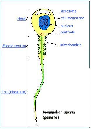 "Semen analysis including sperm morphology assessment has been suggested to be a useful indication of the factors in man's macro-environment, which can modulate or damage spermatogenesis (Mac Leod & Gold 1953). The present study was aimed to determine the reproductive toxic effects of male rat after ingestion of NaF [4.5-9 ppm] through drinking water. The route chosen in this study for exposure was via drinking water to mimic human exposure and to reflect the impact on fertility, after chronic ingestion. The decreased sperm number and motility observed in experimental rats might be responsible for decreasing male fertility. Decrease in male reproductive potential was observed in rats and rabbits after exposure to fluoride (Kumar & Susheela 1994, 1995; Narayana & Chinoy 1994; Zhang et al. 2000; Collins et al. 2001). Besides decreased sperm count, sperm motility, the sperm viability and HOS sperm coiling percentages were also adversely affected in NaF-exposed rats. These changes were greater in rats exposed to higher dose of NaF. The decreased testicular steroidogenic enzyme activity levels may lead to decreased steroidogenesis in experimental rats, which in turn may suppress the reproductive activities in the male rats."
SOURCE: Pushpalatha T, Srinivas M, Sreenivasula Reddy P. (2005). Exposure to high fluoride concentration in drinking water will affect spermatogenesis and steroidogenesis in male albino rats. Biometals 18:207-12.
"The content of NaF in testis and the ratio of apoptotic spermatogenic cell in fluoride treatment groups significantly increased with increased experimental dosage and prolonged experimental period (P < 0.05). Meanwhile, the serum estradiol level significantly decreased (P < 0.05), which was negatively correlated with the content of NaF in testis as well as the ratio of apoptotic spermatogenic cell (P < 0.05). CONCLUSION: Excessive fluoride could lead disturbance to serum estradiol level during some range of dose and time, which is an important factor to spermatogenic cell apoptosis."
SOURCE: Jiang CX, et al. (2005). [Relationship between spermatogenic cell apoptosis and serum estradiol level in rats exposed to fluoride]. Wei Sheng Yan Jiu . 34:32-4.
SOURCE: Krasowska A, et al. (2004). Zinc protection from fluoride-induced testicular injury in the bank vole (Clethrionomys glareolus). Toxicology Letters 147: 229-235.
"This study examined the effect of sodium fluoride, a water pollutant important through the world, including India, on testicular steroidogenic and gametogenic activities in relation to testicular oxidative stress in rats. Sodium fluoride treatment at 20mg/kg/day for 29 days by oral gavage resulted in significant diminution in the relative wet weight of the testis, prostate, and seminal vesicle without alteration in the body weight gain. Testicular delta(5),3beta-hydroxysteroid dehydrogenase (HSD) and 17beta-HSD activities were decreased significantly along with significant diminution in plasma levels of testosterone in the fluoride-exposed group compared to the control. Epididymal sperm count was decreased significantly in the fluoride-treated group and qualitative examination of testicular sections revealed fewer mature luminal spermatozoa in comparison to the control. The seminiferous tubules were dilated in treated animals. Fluoride treatment was associated with oxidative stress as indicated by an increased level of conjugated dienes in the testis, epididymis, and epididymal sperm pellet with respect to control. Peroxidase and catalase activities in the sperm pellet were decreased significantly in comparison to the control. The results of this experiment indicate that fluoride at a dose encountered in drinking water in contaminated areas exerts an adverse effect on the male reproductive system and this effect is associated with indicators of oxidative stress."
SOURCE: Ghosh D, et al. (2002). Testicular toxicity in sodium fluoride treated rats: association with oxidative stress. Reproductive Toxicolology 16(4):385.
"To study the mechanisms of the antagonistic action of selenite on fluoride-induced male reproductive damages , and find out the optimal level of selenite in drinking water against fluoride toxicity... Results: Fluoride could cause the elevation of fluorine concentrations in blood and urine, the abnormalities of trace elements in serum and testis, as well as the significant increase of lipid peroxide (LPO) levels, and the obvious decreases of activities of glutathione peroxidase (GSH-Px) and ATPase in testis and epididymis of rats exposed to fluoride in drinking water (68 mg/L)."
SOURCE: Yang KD, et al. (2002). [Study on antagonistic effects of selenite on fluoride-induced impairments of testis and epididymis in rats]. Chung-Kuo Kung Kung Wei Sheng 18: 427-9.
"The activities of androgen-dependent enzymesacid phosphatase (ACP), lactate dehydrogenase (LDH), and gamma-glutamyl transferase (y-GT-10S)decreased significantly when the ejaculate was treated with NaF at concentrations of 20, 100, 200 µmol/L (0.38; 1.9; 3.8 ppm F-), but they returned to the initial value of the control at 0.1 mol/L (1900 ppm F-)... These changes undoubtedly affect the physiological functions of the sperm."
SOURCE: Zakrzewska H, et al. (2002). In vitro influence of sodium fluoride on ram semen quality and enzyme activities. Fluoride 35: 153-160.
"From the foregoing data, it is evident that the administration of sodium fluoride or aluminium chloride alone induced (reproductive) toxicity in female mice. This toxicity was enhanced by their combined treatment (Group IV) in affecting steroidogenesis in ovary, carbohydrate metabolism in uterus, and causing a hypercholesterolemic effect in mice."
SOURCE: Chinoy NJ, Patel TN. (2001). Efects of sodium fluoride and aluminium chloride on ovary and uterus of mice and their reversal by some antidotes. Fluoride 34: 9-20.
"The effects of sodium fluoride (NaF) ingestion (10 mg NaF/kg body weight) and the possible therapeutic effects of ascorbic acid (AA, 15 mg/animal/day) and/or calcium phosphate (Ca, 25 mg/animal/day) on the reproductive functions and fertility of male mice were investigated. NaF-ingestion brought about a significant decline in sperm acrosomal acrosin and hyaluronidase. Cauda epididymal sperm stained with alcoholic acidic silver nitrate reagent revealed acrosomal damage and deflagellation. However, sperm nuclear integrity was not affected by the treatment. The reduced activity of the enzymes as well as the structural and metabolic alterations in the sperm led to a significant decrease in sperm count, and motility and live:dead ratios but an increase in abnormal sperm which ultimately lead to a poor fertility rate. The cessation of NaF-treatment was not conducive to bringing about a complete recovery. However, the administration of AA or Ca to NaF-treated mice revealed significant recovery from fluoride toxicity in all the above parameters."
SOURCE: Chinoy NJ, Sharma A. (2000). Reversal of fluoride-induced alteration in cauda epididymal spermatozoa and fertility impairment in male mice. Environmental Sciences 7: 29-38.
"Sexually mature male Swiss mice were exposed at 60 days of age to 100, 200 and 300 ppm sodium fluoride (NaF) in their drinking water for 4 weeks or 10 weeks. The effect of NaF exposure on fertility was assessed by breeding these males with untreated female mice after the exposure periods. Fertility was significantly reduced at all three concentrations by exposure for 10 weeks but not for 4 weeks. The number of implantation sites and viable fetuses was significantly reduced in females mated with males that had ingested NaF at a concentration of 200 ppm for 10 weeks. Relative weights of seminal vesicles and preputial glands were significantly increased in mice exposed to 200 and 300 ppm NaF for 4 weeks but not in mice exposed for 10 weeks. These results indicate that long-term ingestion of NaF adversely affects fertility in male mice."
SOURCE: Elbetieha A, et al. (2000). Fertility effects of sodium fluoride in male mice. Fluoride 33: 128-134.
"In summary, we found that sodium fluoride administered in drinking water to rats for 30 days at doses averaging 22.6 mg/kg/day caused definite fetotoxic effects. There was a reduction in the number of viable fetuses and an increase in the number of pregnant rats with resorptions as well as an increase in the total number of resorptions."
SOURCE: Hiyasat AS. (2000). Reproductive Toxic effects of ingestion of sodium fluoride in female rats. Fluoride 33(2): 79-84.
"These results clearly indicate that protein supplementation is beneficial to overcome the toxic effects of fluoride on testicular steroidogenesis, protein, carbohydrate, and energy and oxidation metabolisms in the reporductive organs of male mice. Protein deficiency, on the other hand, aggravates fluoride toxicity. A protein-supplemented diet might therefore substantially mitigate certain fluoride-induced health hazards in humans living in endemic areas."
SOURCE: Chinoy NJ, Mehta D. (1999). Effects of protein supplementation and deficiency on fluoride-induced toxicity in reproductive organs of male mice. Fluoride 32: 204-214.
"Studies on the beneficial effects of vitamins E and D supplementation on functions of caput and cauda epididymides, their spermatozoa, vas deferens and seminal vesicle of sodium fluoride (NaF) treated male mice were carried out. The NaF treatment resulted in significant decrease in the body and epididymis weight but those of vas deferens and seminal vesicle were not affected. NaF treatment brought about alterations in epididymal milieu as elucidated by the significant decrease in levels of sialic acid and protein as well as activity of ATPase in epididymides. As a result, the sperm maturation process was affected leading to a significant decline in cauda epididymal sperm motility and viability. This caused a significant reduction in fertility rate. The cauda epididymal sperm count was also significantly reduced. The data obtained suggest that fluoride treatment induced significant metabolic alterations in the epididymides, vas deferens and seminal vesicles of mice."
SOURCE: Chinoy NJ, Sharma A. (1998). Amelioration of fluoride toxicity by vitamin E and D in reproductive functions of male mice. Fluoride 31: 203-216.
"Conclusions: The modification of some parameters related to fertility by the repeated oral NaF intake, in rodents, suggest that NaF has potential to disturb male fertility."
SOURCE: Pinto R, et al. (1998). NaF may disturb male fertility in rodents. Toxicology Letters 95(Suppl 1): 214.
"Effects of sodium fluoride (NaF) (30 mg kg-1 body weight) and ascorbic acid ingestion along with sodium fluoride for 30 days each were studied to evaluate its possible role as an ameliorative agent on functions of reproductive organs and spermatozoa of the fluorotic guinea pig. The cauda epididymal spermatozoa were highly sensitive to the effects of sodium fluoride as their structural and metabolic alterations led to marked decreases in their motility, live:dead ratio and sperm mitochondrial activity index but increases in sperm abnormalities and alterations in sperm membrane phospholipids, particularly phosphatidylinositol and phosphatidyl serine. The activities of ATPase and succinate dehydrogenase as well as glutathione levels were decreased in testis by sodium fluoride treatment, revealing disturbances in its metabolism."
SOURCE: Chinoy NJ, et al. (1997). Fluoride toxicity in the testis and cauda epididymis of guinea pig and reversal by ascorbate. Medical Science Research 25: 97-100.
"The toxic effects were evaluated of sodium fluoride (NaF) ingestion on the physiology of tissue components of testis and epididymis of adult, male albino rats, and the possible reversal of the effects by use of some antidotes. The results revealed that the testis and cauda epididymal proteins were altered, with disappearance of some proteins and induction of some new ones. This is the first report of such changes... On comparing the alterations in protein profile, phospholipds and glutathione in both tissues, it was evident that the protein profile was disturbed more in testis than in cauda epididymis, whereas phospholipids and gluthathione levels were affected more in cauda than in testis... As the proteins of testis and cauda epididymis are known to be involved as androgen carrier proteins, in testicular functions and in sperm motility, it follows that NaF treatment might affect the levels of these proteins as well as alter sperm motility and viability."
SOURCE: Chinoy NJ, et al. (1997). Fluoride toxicity on rat testis and cauda epididymal tissue components and its reversal. Fluoride 30: 41-50.
"The section on the effects of fluoride on the physiological signs of sexual maturity in the gerbil was a preliminary, pilot study. There were not enough subjects to make any firm conclusions so an interpretation of the data is conjectural. However, the results do suggest that the HF (High-Fluoride) females had an accelerated onset of puberty as judged by several indices of pubertal development in rodents . At 7 weeks, the HF females were significantly heavier than the LF females (p < 0.004); as heavy as the HF males and LF males. The ventral gland in the HF female developed significantly earlier than in the LF female (p < 0.004). Vaginal opening occurred earlier in the HF female than in the LF female (p <0.03)."
SOURCE: Luke J. (1997). The Effect of Fluoride on the Physiology of the Pineal Gland . Ph.D. Thesis. Unive rsity of Surrey, Guildford. p. 173-174.
"At 16 weeks, the HF [High-Fluoride] males had a significantly lower mean testes weight than the LF [Low-Fluoride] males: 1.10 ± 0.11 vs. 1.32 ± 0.18 g, respectively (p <0.002). The reason for this is not clear."
SOURCE: Luke J. (1997). The Effect of Fluoride on the Physiology of the Pineal Gland . Ph.D. Thesis. University of Surrey, Guildford. p. 177.
"The present communication addresses the effect of chronic fluoride toxicity on the structure of rabbit Leydig cells using light, scanning and transmission electron microscopy... [T]he extensive degenerative changes (which are progressive) seen in the Leydig cells due to fluoride toxicity may lead to a decrease in testosterone production resulting initially in regression of seminiferous tubules and structural damage of the epididymis and finally cessation of spermatogenesis."
SOURCE: Susheela AK, Kumar A. (1997). Ultrastructural studies on the leydig cells of rabbits exposed to chronic fluoride toxicity. Environmental Sciences 5:79-94.
"Sodium fluoride (NaF) at a dose of 10 mg/kg body weight was administered orally to male rats (Rettus norvegicus) daily for 30 and 50 days to evaluate the effect of the physiology of some sex accessory glands and sperm functions. The effects of withdrawal upon cessation of NaF ingestion, and of administering ascorbic acid (AA) and/or calcium (Ca++) along with NaF, were also investigated. The results revealed that the NaF treatment caused a significant elevation in serum fluoride levels with a simultaneous rise in Ca++ levels. This could be attributed to the formation of a calcium fluoride complex leading to calcium accumulation. The treatment resulted in structural and metabolic alterations in sperm, leading to low sperm motility, a low sperm mitochondrial activity index (SMAI), reduced viability (live:dead ratio), and changes in sperm membrane phospholipids (particularly phosphatidylinositol, phosphatidylserine and phosphatidylethanolamine, which would affect hormone receptor interaction and their functions). A significant reduction in electrolyte levels of sperm also occurred which would also affect their viability. The protein levels in cauda epididymal sperm suspension, vas deferens, seminal vesicle and prostate were significantly decreased after NaF administration, which may be due to altered protein metabolism by interference of fluoride ions. The changes in epididymal protein profile, with absence of some proteins and induction of some new ones, were probably a result of the "stress proteins" in NaF-treated rats affecting the structural and functional integrity of sperm. Glycogen accumulation in vas deferens and a decrease in fructose in seminal vesicles and vas deferens indicated disturbances in carbohydrate metabolism in these organs. However, withdrawl of treatment resulted in partial recovery. A significant recovery from NaF-induced toxic effects occurred following administation of ascorbic acid and/or calcium, while combined treatment (AA + Ca++) for 70 days manifested a synergistic effect. The transient fluoride-induced effects were reversible. The results, corroborated by earlier data from our laboratory, show that fluoride has a definite effect on male reproduction and fertility. Ascorbic acid and calcium are proposed as therapeutic agents in endemic populations for ameiloration of effects of fluoride."
SOURCE: Chinoy NF, et al. (1995). Amelioration of fluoride toxicity in some accessory reproductive glands and spermatozoa of rat. Fluoride 28: 75-86.
"Fifty four Wistar male rats were randomly divided into three groups, drinking water containing 0.6 mg/L (control group), 100 mg/L, and 200 mg/L sodium fluoride, respectively. Rats were killed at the second, fourth and sixth weeks after experiment initiation, respectively. The levels of serum testosterone, testis cholesterol, and hepatic tissue cholesterol were determined. Results showed that the serum testosterone level had decreased with time in rats drinking water containing 100 and 200 mg/L fluoride. While testis cholesterol level did not change, it was significantly decreased in the liver at the fourth and sixth week when compared with the control group. Results suggest that fluoride may have some harmful effects on the reproductive system in male rats."
SOURCE: Zhao ZL, et al. (1995). The influence of fluoride on the content of testosterone and cholesterol in rat. Fluoride 28: 128-130.
SOURCE: Chinoy NF, et al. (1994). Beneficial effects of ascorbic acid and calcium on reproductive functions of sodium fluoride-treated prepubertal male rats. Fluoride 27: 67-75.
"OBJECTIVE--To address the role of fluoride in causing defects to spermatids and epididymal spermatozoa. METHODS--Male rabbits were treated with 10 mg NaF/kg body weight daily for 18 months and maintained under identical laboratory conditions along with the control rabbits not given NaF. Testis and epididymis (caput) were investigated for ultrastructural details of spermatids and spermatozoa. RESULTS--A wide variety of structural defects were observed in the flagellum, the acrosome, and the nucleus of the spermatids and epididymal spermatozoa of fluoride-treated rabbits. Abnormalities included absence of outer microtubules, complete absence of axonemes, structural and numeric aberrations of outer dense fibers, breakdown of the fibrous sheath, and structural defects in the mitochondria of the middle piece of the flagellum. Detachment and peeling off of the acrosome from the flat surfaces of the nucleus were also observed. CONCLUSION-- The abnormalities observed render the sperm nonfunctional and ineffective, and thus there is a possible role of fluoride in causing infertility."
SOURCE: Kumar A, Susheela AK. (1994). Ultrastructural studies of spermiogenesis in rabbit exposed to chronic fluoride toxicity. International Journal of Fertility and Menopausal Studies 39(3):164-71.
"The effects of ingestion of sodium fluoride (NaF), 10 mg/kg body weight for 50 days, on the structure and metabolism of sperm of albino rats (Rattus norvegicus), were investigated. In different groups of rats, the reversible effects upon withdrawal of NaF treatment and by administering some therapeutic agents, viz., ascorbic acid and calcium alone and in combination with NaF (50 and 70 days), on sperm structure and metabolism were also studied. The results revealed that the sperm acrosomal hyaluronidase and acrosin were reduced after 50 days of NaF treatment. Sperm stained with acidic alcoholic silver nitrate revealed acrosomal damage and deflagellation, which might be causative factors for the reduced activity of the enzymes. These alterations also resulted in a decline in sperm motility. The cauda epididymal sperm count was decreased, perhaps because of spermatogenic arrest. Thus, the low sperm motility and count ultimately contributed toward reduction in fertility by NaF treatment. However, withdrawal of NaF treatment for 70 days produced incomplete recovery, while administration of ascorbic acid and calcium, individually and in combination, brought about significant recovery of fluoride-induced effects. Thus, the effects of fluoride on sperm structure and metabolism of rats are transient and reversible."
SOURCE: Narayana MV, Chinoy NJ. (1994). Reversible effects of sodium fluoride ingestion on spermatozoa of the rat. International Journal of Fertility and Menopausal Studies 39(6):337-46.
"In view of reports of infertility among human populations in fluorosis prevailing regions, we investigated the effect of fluoride ingestion on testicular steroidogenesis in rats. Sodium fluoride (NaF) was administered to the rats orally at a daily dose of 10mg/kg bodyweight for 50 days. The treatment did not cause significant change in testicular cholesterol levels, indicating that metabolism was not altered and that there was no hypo/hypercholesterolemic effect. In addition, activities of the intermediary enzymes in androgenesis, viz., 3ß- and 17ß-hydroxysteroid dehydrogenase were only modestly decreased by NaF ingestion. Subsequently, the determination of circulating androgen levels in NaF-treated rats showed a downward trend compared to those of the control group, suggesting alteration in testosterone concentration. The histomorphometric studies revealed significant change in the Leydig cell diameter m correlation with the androgen levels. These results indicate that fluoride does interfere with steroidogenesis in short-term lowdose exposures in rats."
SOURCE: Narayana MV, Chinoy NJ. (1994). Effect of fluoride on rat testicular steroidogenesis. Fluoride 27: 7-12.
"In fluorotic rats, testicular cholesterol and serum testosterone levels were not affected. However, succinate dehydrogenase activity in testis was inhibited. Similarly, adenosine triphosphatase activity and sialic acid levels in epididymides were also suppressed with more pronounced effect on cauda epididymis. Consequently, sperm motility and count were decreased leading to a significant decline in fertility by fluoride treatment. Hence, rat is also sensitive to fluoride toxicity."
SOURCE: Chinoy NJ, et al. (1992). Effects of fluoride ingestion on the physiology of reproductive organs of male rats. Journal of Environmental Biology 13: 55-61.
"Summary: Sodium fluoride (NaF) fed to adult male albino mice at a dose of 10 mg and 20 mg/kg body weight, caused a significant decrease in sperm county and motility. Scanning electron microscopy and silver nitrte staining showed large numbers of deflagellated spermatozoa, with acrosomal, midpiece and tail abnormalities. The treatment caused loss of fertility rate when normal cycling female mice were mated with treated males."
SOURCE: Chinoy NJ , Sequeira E. (1992). Reversible fluoride induced fertility impairment in male mice. Fluoride 25 71-76.
"Male Wistar rats were exposed to fluoride (F) at concentrations of 100- and 200 ppm in their drinking water for 6- and 16 weeks. The high F intake caused several-fold increase in the F concentrations in the testes and bone as compared with control rats, both after the 6- and 16 wk exposure; the bone F, but not testicular F, appeared to increase with dose and time. F exposure (100- and 200 ppm) decreased significantly the concentrations of zinc (Zn) in the testes, plasma, liver and kidneys particularly in the 16 wk groups; in the bone Zn tended to increase, however... Fifty percent of the 100- and 200 ppm F rats after 16 weeks exhibited histopathologic changes in the germinal epithelium of the testes, which resembled those in Zn-deficient rats. The data suggest that a deprivation of testicular Zn due to a high F intake may be directly responsible for the injury of testicular tubules."
SOURCE: Krasowska A, Wlostowski T. (1992). The effect of high fluoride intake on tissue trace elements and histology of testicular tubules in the rat. Comparative Biochemistry and Physiology: Part C 103(1):31-4.
"A single microdose (50 micrograms/50 microL) injection of sodium fluoride (NaF) into the vasa deferentia of adult male albino rats (Rattus norvegicus) caused arrest of spermatogenesis and absence of spermatozoa in the lumina of the seminiferous tubules of the testes, which consequently led to a decline in the sperm count in the caudae epididymides. Scanning electron microscopy of cauda and vas deferens sperm revealed deflagellation and tail abnormalities. This is probably related to the alterations in the internal milieu of these organs which rendered the spermatozoa immotile and consequently caused fertility impairment in the experimental animals. Thus microdoses of sodium fluoride were found to affect reproductive function and fertility rate."
SOURCE: Chinoy NJ, et al. (1991). Microdose vasal injection of sodium fluoride in the rat. Reproductive Toxicolology 5(6):505-12.
"Fluoride was orally administered to rabbits at 10 mg NaF/kg body weight for 18 or 29 months. The animals were then killed and the structure of the testis, epididymis and vas deferens studied under light and scanning electron microscopes. In animals treated for 29 months, the spermatogenic cells in the seminiferous tubules were disrupted, degenerated and devoid of spermatozoa. In animals treated for 18 or 29 months, loss of cilia on the epithelial cells lining the lumen of the ductuli efferentes of the caput epididymidis and of stereocilia on the epithelial cells lining the lumen of the vas deferens was observed. In some regions of the epithelial lining of the lumen of the ductuli efferentes and vas deferens, the boundaries of the cells were not clear and appeared to be peeled off. Mucus droplets were abundant in the vas deferens of control animals, but absent in both the treated groups. Spermatogenesis ceased only in animals treated for 29 months. The difference in the structural changes observed in the testes of the 2 treated groups may have been due to the blood-testis barrier. It is concluded that ingestion of high concentrations of fluoride has harmful effects on the male reproductive system."
SOURCE: Susheela AK, Kumar A. (1991). A study of the effect of high concentrations of fluoride on the reproductive organs of male rabbits, using light and scanning electron microscopy. Journal of Reproductive Fertility 92(2):353-60.
"The aim of the study was to evaluate relationship between infertility and the histological structure of the testes following the subcutaneous administration of different doses of sodium fluoride (5, 10, 20 and 50 mg/kg/day), for 100 days, to groups of six male albino rabbits; the six control animals were given 1 cc distilled water/kg b.w./day for the same length of time. Deficient maturation and differentiation of the spermatocytes and an increase in the amount of interstitial tissue were found in the experimental animals. In the higher dosage groups, spermatogenesis stopped and the seminiferous tubules became necrotic. The study thus established the existence of a definite relationship between fluorosis and testicular damage."
SOURCE: Shashi A. (1990). Histopathological changes in rabbit testes during experimental fluorosis. Folia Morphol (Praha) 38(1):63-5.
"Albino rabbits were injected sodium fluoride solutions in the concentration of 5, 10, 20 and 50 mg/kg body weight/day subcutaneously for 100 days. The control rabbits were given 1 cc of distilled water for the same period and sacrificed. The ovary was examined for histopathological changes. Animals in control and 5 mg fluoride treated groups displayed normal follicles with oocytes and interstitial tissue in ovaries. In animals treated with 10 and 20 mg fluoride, ovary exhibited congested oocytes in the follicles, necrosis of follicle cells and interstitial oedema. The degenerative changes were most pronounced in animals treated with 50 mg fluoride, in which complete atrophy of follicles along with oocyte disintegration and marked necrosis of cells accompanied by infiltration of monocytes, lymphocytes and histiocytes in interstitial tissue occurred. The data indicate that the structural alterations in the ovary were more pronounced with the concomitant increase in the dose of fluoride."
SOURCE: Shashi A. (1990). Histopathological changes in rabbit ovary during experimental fluorosis. Indian Journal of Pathology and Microbiology 33(2):113-7.
"The effect of high fluoride intake (100 and 200 ppm) in the ration was studied in male rats. After sixty days of treatment, rats showed a decrease in the mean diameter of the seminiferous tubules and the percentage of the tubules containing spermatozia and increase in the in the thickness of the peritubular membranes. The effect was more prominent with the higher dose of fluoride. Serum testosterone level in rats [which] received 200 ppm fluoride showed a sharp decrease, whereas in those treated with 100 ppm did not differ significantly from the control. The fertility performance of treated rats was reduced and the results revealed a reduction in the number of pregnant females and newborns of both treated groups. Lowest dose of fluoride had a similar but less marked effect on the fertility performance than the higher dose. It is concluded that the high fluoride intake causes a decline in the reproductive performance of the adult male rats, although the clinical signs in the teeth are absent."
SOURCE: Araibi AA, et al. (1989). Effect of high fluoride on the reproductive performance of the male rat. Journal of Biological Sciences Research 20: 19-30.
"The effects of sodium fluoride (NaF) ingestion in two doses (10 and 20 mg/kg body weight) for 30 days on histology and histocytometry of reproductive organs of the adult male mouse were investigated. In order to study reversibility, treatment was withdrawn for one and two months... NaF treatment caused severe disorganization and denudation of germinal epithelial cells of seminiferous tubules with absence of sperm in the lumina. The Leydig cell and nucleus diameters were not affected. The caput epididymis showed fewer changes than the cauda. However, epithelial cell nuclear pyknosis and absence of luminal sperm were observed. A reduction in epithelial cell height, nuclear pyknosis, denudation of cells, and absence of sperm occurred in the cauda epididymis. The vas deferens epithelium showed nuclear pyknosis, clumped stereocilia, and cell debris but no sperm in the lumen and an increase in the lamina propria. The prostate and seminal vesicles were not affected by treatment. Withdrawal of treatment caused marked recovery in the histoarchitecture of these organs. The effects of NaF treatment are therefore transient and reversible."
SOURCE: Chinoy NJ , Sequeira E. (1989). Effects of fluoride on the histoarchitecture of reproductive organs of the male mouse. Reproductive Toxicolology 3(4):261-7.
"Reduction of ingested fluoride in a skulk of silver foxes resulted in the reduction of fluoride burden, decreased neonatal mortality and increased kit production during a two breeding and whelping season period."
SOURCE: Eckerlin RH, et al. (1988). Ameliorative effects of reduced food-borne fluoride on reproduction in silver foxes. Cornell Veterinarian 78(4):385-91.
"Sixty-six eastern screech-owls (Otus asio) were paired and randomly assigned to dietary treatment groups of 0, 40, or 200 ppm (mg/kg) fluoride (as sodium fluoride) in November 1981. Hatching success was adversely affected at the 200 ppm (mg/kg) level, suggesting potential detrimental impacts to wild populations exposed to fluoride pollution."
SOURCE: Pattee OH, et al. (1988). Effects of dietary fluoride on reproduction in Eastern Screech-Owls. Archives of Environmental Contamination and Toxicology 17: 213-218.
"Genotoxicity of Sodium fluoride was evaluated in mice in vivo with the help of different cytogenetic assays. The frequency of chromosome aberration was dose - and time - dependent but not exactly route-dependent. Fractionated dosing induced less aberration. Incidence of micronucleus and sperm abnormality increased with dose. Relative sensitivity of the three assays has been found to be: Sperm abnormality > Chromosome aberration > Micronucleus. The present results have revealed the mutagenic property of NaF."
SOURCE: Pati PC, Bhunya SP. (1987). Genotoxic effect of an environmental pollutant, sodium fluoride, in mammalian in vivo test system. Caryologia 40:79-87.
"The results provide unequivocal evidence that 250 uM fluoride inhibits testosterone secretion by rat testes perfused in vitro. Previous investigators have reported that 5-10 mM fluoride stimulates adenylate cyclase, inhibits metabolic reactions, and inhbits testosterone biosynthesis. The present observation of deleterious effects by 250 uM fluoride (5 ppm) emphasizes the sensitivity of steroidogenesis to fluoride."
SOURCE: Chubb C. (1985). Reproductive toxicity of fluoride. Journal of Andrology 6: 59.
"The effects on reproduction in screech owls (Otus asio) of chronic dietary sodium fluoride administration at 0, 40, and 200 ppm were examined. Fluoride at 40 ppm resulted in a significantly smaller egg volume, while 200 ppm also resulted in lower egg weights and lengths. Day-one hatchlings in the 200 ppm group weighed almost 10% less than controls and had shorter crown-rump lengths... T hese results, in combination with the findings of Pattee et al., revealed significant impairment of overall reproduction, suggesting that sodium fluoride could cause slight to moderate reproduction disorders in owls in fluoride-polluted areas."
SOURCE: Hoffman DJ, et al. (1985). Effects of fluoride on screech owl reproduction: teratological evaluation, growth, and blood chemistry in hatchlings. Toxicology Letters 26(1):19-24.
"A marked fall (p <0.01) in the testosterone production was recorded at a fluoride concentration of 100 ppm and testosterone synthesis was maximally inhibited (p <0.01) at 200 ppm. There was a noticeable, though marginal, inhibition in testosterone synthesis even at 10 ppm fluoride concentration. From 1 ppm to 200 ppm, the degree of inhibition of testosterone synthesis seems to be dependent on fluoride concentration."
SOURCE: Kanwar KC, Vig PS, Kalla NR (1983). In vitro inhibition of testosterone synthesis in the presence of fluoride ions. IRCS Medical Science 11: 813-814.
"The study was designed in order to assess the relationship between infertility and histological structure of testes following administration of varying doses of sodium fluoride. One hundred adult male albino mice were fed 10 ppm (Group A), 500 ppm (Group B) and 1000 ppm (Group C) of sodium fluoride in drinking water. The Group A animals were sacrificed at the end of one month, Group B after two and Group C after three months. The testes were removed and, after being processed in the usual manner, they were stained with hematoxylin and eosin. In Groups B and C, the higher dosage groups, there was a lack of maturation and differentiation of spermatocytes. In animals sacrificed at the end of three months, spermatogenesis had stopped and the seminiferous tubules had become necrotic. A definite relationship between fluorosis and damage to the testes has, therefore, been established by this study."
SOURCE: Kour K, Singh J. (1980). Histological finding of mice testes following fluoride ingestion. Fluoride 13: 160-162.
SOURCE: van Rensburg SWJ, de Vos WH. (1966). The influence of excess fluorine intake in the drinking water on reproductive efficiency in bovines. The Onderstepoort Journal of Veterinary Research 33: 185-194.