HEALTH
EFFECTS: Biology of Dental Fluorosis
Key Findings -
The Biology of Dental Fluorosis:
Excessive ingestion of fluoride during the early childhood years
may result in a disorder of the teeth known as dental
fluorosis. Teeth with fluorosis have an
increase in porosity in the subsurface enamel ("hypomineralization").
The increased porosity of enamel found in fluorosis is a result
of a fluoride-induced impairment in the clearance of proteins
(amelogenins) from the developing teeth.
Despite over 50 years of research, the exact mechanism by which
fluoride impairs amelogin removal is not yet fully
understood. It is believed, however, that it involves a fluoride-induced
toxic effect on the cells (ameloblasts) involved in enamel
formation.
In addition to affecting the enamel, dental fluorosis may also
affect the underlying dentin as well, thus
suggesting that "fluoride may exert
effects at the cellular level well beyond tooth
development."
Notable Quotes -
The Biology of Dental Fluorosis:
"The fact that human dentin also exhibits
hypomineralization in human fluorotic teeth
indicates that fluoride exerts its effects on very basic processes
involved in biomineralization in general, irrespective
of whether crystal formation and growth occurs in mesenchymally
or ectodermally derived mineralized tissues.
However, relatively little work has been done to identify the
mechanisms by which low serum levels of fluoride which result
in dental fluorosis affect the development of mineralizing tissues."
SOURCE: Fejerskov O, Richards A, DenBesten P. (1996). The effect
of fluoride on tooth mineralization. In: Fejerskov O, Ekstrand
J, Burt B, Eds. Fluoride in Dentistry,
2nd Edition. Munksgaard, Copenhagen. pp. 112-152.
"it is illogical to assume that tooth
enamel is the only tissue affected by low daily doses of fluoride
ingestion."
SOURCE: Dr. Hardy Limeback, Head of Preventive Dentistry, University
of Toronto. (2000). Why I am
now Officially Opposed to Adding Fluoride to Drinking Water.
"Like bones, a child's teeth are alive
and growing. Flourosis is the result of fluoride rearranging the
crystalline structure of a tooth's enamel as it is still growing.
It is evidence of fluoride's potency and ability to cause physiologic
changes within the body, and raises concerns about similar damage
that may be occurring in the bones."
SOURCE: Environmental Working Group, "National
Academy Calls for Lowering Fluoride Limits in Tap Water",
March 22, 2006.
"A linear correlation between the Dean
index of dental fluorosis and the frequency of bone fractures
was observed among both children and adults."
SOURCE: Alarcon-Herrera
MT, et al. (2001). Well Water Fluoride, Dental fluorosis, Bone
Fractures in the Guadiana Valley of Mexico. Fluoride 34(2):
139-149.
"It seems prudent at present to assume
that the ameloblasts are not the only cells in the body whose
function may be disturbed by the physiological concentrations
of fluoride which result from drinking water containing 1 ppm."
SOURCE: Groth, E. (1973), Two
Issues of Science and Public Policy: Air Pollution Control in
the San Francisco Bay Area, and Fluoridation of Community Water
Supplies. Ph.D.
Dissertation, Department of Biological Sciences, Stanford University,
May 1973.
"The safety of the use of fluorides ultimately
rests on the assumption that the developing enamel organ is most
sensitive to the toxic effects of fluoride. The results from this
study suggest that the pinealocytes
may be as susceptible to fluoride as the developing enamel organ."
SOURCE: Luke J. (1997). The
Effect of Fluoride on the Physiology of the Pineal Gland.
Ph.D. Thesis. University of Surrey, Guildford. p. 176
Biology of Dental Fluorosis
- Increased Porosity due to Impaired
Clearance of Amelogenins: (back
to top)
"Fluorosis is a hypomineralization of
enamel caused by the retention of amelogenin
proteins by fluoride. The affected enamel does not mature
and has surface and subsurface porosities."
SOURCE: Allen K, et al. (2004). Using microabrasive material to
remove fluorosis stains. Journal
of the American Dental Association
135:319-23.
"Dental or enamel fluorosis occurs when excess amounts of
fluoride are ingested during tooth development (1-8 years of age).
It is characterized by increased porosity (or
hypomineralization) of the subsurface enamel and well mineralized
surface layer of enamel. Mildly fluorosed enamel is fully functional,
but may be cosmetically objectionable. As the
severity of dental fluorosis increases, the depth of the enamel
involvement and the degree of porosity increases. More
severely fluorosed enamel is more porous, pitted, and discolored
and is prone to fracture and wear because the well mineralized
zone is very fragile to mechanical stress."
SOURCE: Agency for Toxic Substances & Disease Registry [ATSDR].
(2003). Toxicological profile
for Fluorides, Hydrogen Fluoride, and Fluorine.
Atlanta, GA: U.S. Department of Health and Human Services, Public
Health Service.
"The observations that the fluorosed enamel
retains a relatively high proportion of immature matrix proteins,
characterized by high proline contents, support the notion of
an incomplete removal of amelogenin proteins under excessive fluoride
ingestion during development. Whitford (1997) recently stated
that "although several other fluoride-induced effects might
be involved in the aetiology of fluorosis, it now appears that
inhibition of enzymatic degradation of amelogenins, which
may delay their removal from the developing enamel and impair
crystal growth, may be of critical importance".
SOURCE: Aoba T, Fejerskov O. (2002). Dental
fluorosis: chemistry and biology. Critical
Reviews of Oral Biology and Medicine
13: 155-70.
"Enamel fluorosis results from increased
porosity in the formed enamel, probably due to a
delay in the removal of amelogenin proteins during enamel
formation. The hydrolysis and removal of amelogenin
from the enamel matrix is critical for tooth growth and development.."
SOURCE: DenBesten PK, et al. (2002). Effects of fluoride on rat
dental enamel matrix proteinases. Archives
of Oral Biology 47: 763-770.
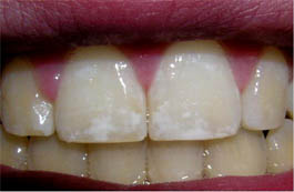
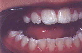
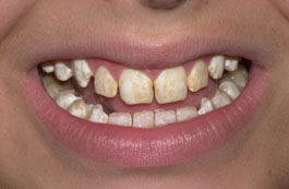
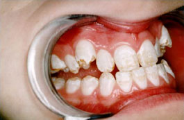
"Dental fluorosis is characterized by
an increasing porosity (hypomineralization) of the subsurface
enamel, causing the enamel to appear
opaque. The clinical features
include changes ranging from barely discernible fine white lines
running across the teeth to entirely chalky white teeth. In advanced
stages, the enamel may become so porous that the outer layers
break down and the exposed porous subsurface becomes discolored."
SOURCE: Fomon SJ, et al. (2000). Fluoride
intake and prevalence of dental fluorosis: trends in fluoride
intake with special attention to infants. Journal
of Public Health Dentistry
60: 131-9.
"Fluorosed enamel is characterized by
retention of amelogenins in the early-maturation stage
of enamel and by the formation of subsurface hypomineralization."
SOURCE: Sapov K, et al. (1999). A laboratory assessment of enamel
hypoplasia of teeth with varying severities of dental fluorosis.
Journal of Oral Rehabilitation
26: 672-7.
"Dental fluorosis is defined as a permanent
hypomineralization of enamel, characterized by greater
surface and subsurface porosity than in normal enamel, that results
from excess fluoride (F) reaching the developing tooth during
developmental stages... Excess F available to the enamel during
maturation disrupts mineralization and results in excessive
retention of enamel proteins."
SOURCE: Burt BA ; Eklund SA. (1999). Dentistry, Dental Practice,
and the Community (5th Ed). WB Saunders Co; Philadelphia.
"excessive intake (of fluoride) leads
to dental and skeletal fluorosis characterized by hypomineralization
of the calcified tissues."
SOURCE: Milan AM, et al. (1999). Altered phosphorylation of rat
dentine phosphoproteins by fluoride in vivo. Calcified
Tissue International 64:234-8.
"[T]he hypomineralized regions of
fluorosed enamel might be an arrest of enamel
maturation... In support of this hypothesis was the finding
that human fluorosed enamel, when compared with normal mature
enamel, had a similar total protein content, but the fluorosed
enamel retained a relatively high proportion of immature matrix
proteins.”
SOURCE: Fejerskov O, et al. (1990). The nature and mechanisms
of dental fluorosis in man. Journal
of Dental Research 69(Spec
Iss): 692-700.
"Any use of fluorides, whether systemic or topical, in caries
prevention and treatment in children results in ingestion and
absorption of fluoride into the blood circulation. The mineralization
of teeth under formation may be affected so that dental fluorosis
may occur. Dental fluorosis reflects an increasing
porosity of the surface and subsurface enamel, causing the enamel
to appear opaque. The clinical features represent a continuum
of changes ranging from fine white opaque lines running across
the tooth on all parts of the enamel to entirely chalky white
teeth. In the latter cases, the enamel may be so porous (or hypomineralized)
that the outer enamel breaks apart posteruptively and the exposed
porous subsurface enamel becomes discolored."
SOURCE: Fejerskov O, et al. (1990). The
nature and mechanisms of dental fluorosis in man. Journal
of Dental Research 69(Spec
Iss): 692-700.
“Fluorosed enamel is also characterized
by a delay in the withdrawal of protein when compared with control
enamel."
SOURCE: Denbesten PK, et al. (1985). Changes in the fluoride-induced
modulation of maturation stage ameloblasts of rats. Journal
of Dental Research 64: 1365-70
.
"Enamel maturation has been characterized by the progressive
deposition of mineral and withdrawal of organic matrix and water.
It is evident that high chronic levels of fluoride
interferes with this process."
SOURCE: DenBesten PK,
Crenshaw MA. (1984). The effects of chronic high fluoride levels
on forming enamel in the rat. Archives
of Oral Biology 29:675-9.
Biology of Denal Fluorosis
- Impacts on the Dentin:
(back to top)
"To the authors’ best knowledge, this study is the
first to analyze the correlation between dentin [F] and the mechanical
and structural properties of dentin. Despite previous work on
dentin quality (structural, material and mechanical properties),
no studies have evaluated the relationship between dentin mechanical
and structural properties and tooth [F]. This relationship is
important owing to the increase in F ingestion worldwide (e.g.
halo effect, F in dentifrices and other dental products).
In this study, we showed that enamel F concentration does not
correlate with any of the parameters tested, while
dentin [F] correlates positively
with dentin tubule size and negatively with ultrasound velocity.
Moreover, the severity of DF [Dental
Fluorosis] correlates positively with dentin tubule density and
ultrasound velocity. It is known that ultrasound velocity relates
to elastic modulus, and that dentin tubule size appears to be
related to tooth sensitivity. Dentin sensitivity is believed
to be related to dentin tubule permeability, which in turn is
related to dentin tubule size. Therefore, one can infer that dentin
[F] and/or DF severity, influence dentin elastic modulus and tooth
sensitivity.
When analyzing all teeth together,
our study showed a positive correlation between dentin [F] and
dentin tubule size, demonstrating wider dentin tubules in teeth
with higher levels of F in dentin. This is interesting
because in humans, histological changes caused by the ingestion
of F have more easily been detected in the enamel, but in severe
fluorosis the dentin has also shown histological modifications.
Fluoride concentration has been shown to influence crystal size,
and some evidence indicates that F has an effect on cell function,
either directly through interactions with the developing ameloblasts
and/or odontoblasts or more indirectly by interacting with the
extracellular matrix. Fluoride has been shown to increase bone
formation and to increase mineralization lag time in bone, increasing
the time between matrix deposition and its mineralization. The
Fluoride content in the tooth structure may therefore have the same type
of action on odontoblast and dentin mineralization (i.e. decreasing
the mineralization rate). A hypomineralized enamel and dentin
is therefore formed. This action would
then explain the positive relationship between dentin tubule size
and tooth [F], where hypomineralization, caused by F concentration,
would create wider dentin tubules. Another possible hypothesis
is that F would influence crystal growth, forming an impaired
dental structure with wider dentin tubules."
SOURCE: Vieira AP, Hancock R, Dumitriu M, Limeback H, Grynpas
MD. (2006). Fluoride's effect on human dentin ultrasound velocity
(elastic modulus) and tubule size. European
Journal of Oral Science 114:83-8.
"Fluoride (F) has been a useful instrument in caries prevention.
However, only limited data exist on the effect of its long-term
use on dentin mineralization patterns and microhardness... Teeth
were analyzed for DF severity, dentin [F], enamel [F], dentin
microhardness, and dentin mineralization. Dentin
[F] correlated with DF severity; enamel [F] correlated with dentin
microhardness and dentin mineralization; DF severity correlated
with dentin microhardness."
SOURCE: Vieira A, Hancock R, Dumitriu M, Schwartz M, Limeback
H, Grynpas M. (2005). How does fluoride affect dentin microhardness
and mineralization? Journal
of Dental Research 84:951-7.
"Although the exact mechanisms are unclear, the
mineralization of connective tissues such as dentine is apparently
altered in the presence of fluoride."
SOURCE: Milan AM, et al. (2001). Fluoride alters casein kinase
II and alkaline phosphatase activity in vitro with potential implications
for dentine mineralization. Archives
of Oral Biology 46:343-51.
"The fact that human dentin also exhibits
hypomineralization in human fluorotic teeth indicates that fluoride
exerts its effects on very basic processes involved in biomineralization
in general, irrespective of whether crystal formation and
growth occurs in mesenchymally or ectodermally derived mineralized
tissues. However, relatively little work
has been done to identify the mechanisms by which low serum levels
of fluoride which result in dental fluorosis affect the development
of mineralizing tissues."
SOURCE: Fejerskov O, Richards A, DenBesten P. (1996). The effect
of fluoride on tooth mineralization. In: Fejerskov O, Ekstrand
J, Burt B, Eds. Fluoride in Dentistry,
2nd Edition. Munksgaard, Copenhagen. pp. 112-152.
"Thus, the present study has demonstrated that exposure
of the dentin-pulp complex to higher concentrations of uoride
inuences the mineralization process in the transition from predentin
to dentin, which may well have a mechanistic basis in the uoride-induced
extracellular matrix changes arising in this region of the tissue.
In view of the continued apposition of dentin throughout life,
these observations indicate that exposure
to high levels of fluoride may exert effects at the cellular level
well beyond tooth development during primary, physiological
secondary and tertiary dentinogenesis. There are also implications
for the regeneration and repair of dentin after injury. The critical
importance of growth factors sequestered within the dentin matrix
in dentin repair and their association with extracellular matrix
components imply that biological repair processes may also be
susceptible to the effects of excessive uoride exposure."
SOURCE: Moseley R, et al. (2003). The influence of fluoride exposure
on dentin mineralization using an in vitro organ culture model.
Calcified Tissue International
73:470-5.
"The sevenfold reduction in phosphate
content of DPP isolated from fluorotic dentine evident in the
present study will undoubtedly have an influence on the anionic
nature of these macromolecules. Such a major change in
the biochemical structure of DPP, together with those previously
reported for other macromolecules such as proteoglycans, are likely
to be important in considering the hypomineralization associated
with fluorosis."
SOURCE: Milan AM, et al. (1999). Altered phosphorylation of rat
dentine phosphoproteins by fluoride in vivo. Calcified
Tissue International 64:234-8.
"In 1925 Beust called attention to the
fact that in adddition to the enamel, the dentin was likewise
affected, a condition which he termed mottled dentin."
SOURCE: Dean HT. (1936). Chronic
endemic dental fluorosis (mottled enamel). Journal
of the American Medical Association
107: 1269-1273.
Biology of Dental Fluorosis
- Mechanisms Not Yet Fully Understood:
(back to top)
"Both collagenous and noncollagenous components appear to
undergo structural alterations during uorosis, although the
precise mechanisms are unclear."
SOURCE: Moseley R, et al. (2003). The
influence of fluoride exposure on dentin mineralization using
an in vitro organ culture model. Calcified
Tissue International 73:470-5.
"While it is well-accepted that fluoride interacts with
mineralized tissues and, at elevated concentrations, disturbs
the mineralization process, the molecular mechanisms
that underlie the pathogenesis of dental fluorosis are not known."
SOURCE: Everett ET, et al. (2002). Dental fluorosis: variability
among different inbred mouse strains. Journal
of Dental Research 81: 794-8.
"In the past, several explanations or
hypotheses have been proposed for the fluoride-induced retention
of amelogenin-derived fragments (as well as the degraded
products of other matrix proteins) in the matured enamel. The
postulated fluoride effects are categorized into two groups: (i)
on intracellular events, including gene expression, synthesis,
trafficking and secretion of proteins, resorption and degradation
of the once-secreted products; and (ii) on extracellular events
constituting multivarious interactions between and among matrix
proteins, proteases, crystals, and other fluid constituents, particularly
fluoride and calcium ions."
SOURCE: Aoba T, Fejerskov O. (2002). Dental
fluorosis: chemistry and biology. Critical
Reviews of Oral Biology and Medicine
13: 155-70.
"Considering the complexity of the biological mineralization
process, the exact mechanism leading to dental
fluorosis is not fully understood."
SOURCE: Susheela AK, Bhatnagar M. (1999). Structural aberrations
in fluorosed human teeth: Biochemical and scanning electron microscopic
studies. Current Science77: 1677-1680.
"The fact that human dentin also exhibits
hypomineralization in human fluorotic teeth indicates that fluoride
exerts its effects on very basic processes involved in biomineralization
in general, irrespective of whether crystal formation and
growth occurs in mesenchymally or ectodermally derived mineralized
tissues. However, relatively little work has
been done to identify the mechanisms by which low serum levels
of fluoride which result in dental fluorosis affect the development
of mineralizing tissues."
SOURCE: Fejerskov O, Richards A, DenBesten P. (1996). The effect
of fluoride on tooth mineralization. In: Fejerskov O, Ekstrand
J, Burt B, Eds. Fluoride in Dentistry,
2nd Edition. Munksgaard, Copenhagen. pp. 112-152.
“Fluoride (F-) that reaches developing teeth induces defects
in the hard tissues, particularly in the enamel. This is
broadly the mechanism of dental fluorosis but many
details of the mechanism, including the exact minimal threshold
doses are not clear.”
SOURCE: Bronckers AL, Woltgens JH. (1985). Short-term effects
of fluoride on biosynthesis of enamel-matrix proteins and dentine
collagens and on mineralization during hamster tooth-germ development
in organ culture. Archives
of Oral Biology 30: 181-91.
"The mechanism underlying the development
of dental fluorosis remains unknown."
SOURCE: Angmar-Mansson B, Whitford GM. (1984). Enamel fluorosis
related to plasma F levels in the rat. Caries Research 18:25-32.
Biology of Dental Fluorosis
- Attempts to Elucidate the Toxic
Effect Underlying Fluorosis: (back
to top)
"In addition to effects on mineral structure and extracellular
processes, NaF affects intracellular pathways leading to alterations
in the actin cytoskeleton. These alterations in 'functional morphology’'
correlate with interference with the Rho pathway, and are expected
to affect ameloblast cyclic morphologic changes known to be disturbed
in fluoride-treated animals. The Rho pathway
in ameloblasts may provide a target for fluoride, potentially
leading also to Rho-linked changes in gene expression. "
LI Y, et al. (2005). Effects of sodium fluoride on the actin cytoskeleton
of murine ameloblasts. Archives
of Oral Biology (in press)
"we have been able to more accurately
localise the pathological effects of fluoride in altering mineralisation
patterns within fluorotic teeth... In summary, the present
study has demonstrated important structural and quantitative changes
in different PG species (decorin, biglycan and versican) within
the individual tissue compartments of the dentine–pulp complex,
following fluoride exposure. Such changes probably
reflect the effects of fluoride on both the synthesis and extracellular
processing of these molecules, the consequences of which will
be to influence the mineralisation process, thereby providing
a pathogenic basis for the altered mineralisation patterns observed
during fluorosis."
SOURCE: Waddington RJ, et al. (2004). Fluoride-induced changes
to proteoglycan structure synthesised within the dentine–pulp
complex in vitro. Biochimica
et Biophysica Acta 1689:142-51.
"We conclude that the ingestion of
fluoride resulting in a serum fluoride
of 5-10 uM (95-190 ppb) can affect the amount of active proteinase
(enzyme) present in maturation-stage enamel in the rat.
In addition, fluoride at concentrations as low
as 2 uM (38 ppb) can reduce metalloproteinase activity at low
pH. These combined effects of fluoride on enamel might
contribute to a mechanism by which high concentrations of systemic
fluoride can affect the hydrolysis of enamel matrix protein and
subsequent biomineralization, resulting in fluorosis."
SOURCE: DenBesten PK, et al. (2002). Effects of fluoride on rat
dental enamel matrix proteinases. Archives
of Oral Biology 47: 763-770.
"Overall these results provide further verification that
fluoride may alter post-translational events
during fluorosis through enzyme activity, and they may
aid in the elucidation of a mechanism for fluorosis."
SOURCE: Milan AM, et al. (2001). Fluoride alters casein kinase
II and alkaline phosphatase activity in vitro with potential implications
for dentine mineralization. Archives
of Oral Biology 46:343-51.
"Of the several mechanisms proposed for
the adverse effect on tooth development, the most likely is that
fluoride has an effect on cell function, either through
interactions with the developing ameloblasts or the intracellular
matrix."
SOURCE: Fomon SJ, et al. (2000). Fluoride
intake and prevalence of dental fluorosis: trends in fluoride
intake with special attention to infants. Journal
of Public Health Dentistry
60: 131-9.
"In our previous studies on the enamel fuorosis model rat,
morphological observation of the secretory
ameloblast shows accumulation of transport vesicles, disorganization
of Golgi stacks and accumulation of abnormal large granules;
pertussis toxin-induced adenosine diphosphate (ADP)-ribosylation
of the membrane fraction reveals activation
of trimeric G proteins bound to rough endoplasmic reticulum
(rER) and Golgi membranes with fluoride treatment. These
findings suggest that fluoride results in aberrant intracellular
transport in the ameloblast through the G proteins."
Matsuo S, et al. (2000). Fluoride-induced ultrastructural changes
in exocrine pancreas cells of rats: fluoride disrupts the export
of zymogens from the rough endoplasmic reticulum (rER).
Archives of Toxicology 73:611-7.
"Such a major change in the biochemical
structure of DPP, together with those previously reported
for other macromolecules such as proteoglycans, are
likely to be important in considering the hypomineralization associated
with fluorosis."
SOURCE: Milan AM, et al. (1999). Altered phosphorylation of rat
dentine phosphoproteins by fluoride in vivo. Calcified
Tissue International 64:234-8.
“An increase in fluoride content
and decrease in calcium content in fluorosed
human teeth were observed when compared to the control.”
SOURCE: Susheela AK, Bhatnagar M. (1999). Structural aberrations
in fluorosed human teeth: Biochemical and scanning electron microscopic
studies. Current Science
77: 1677-1680.
"[S]tructural alterations of ameloblastic
layer result in the retardation of enamel matrix formation
and its mineralization. Calcium deficiency and
generalized malnutrition disturb the physiological conditions
that affect amelogenesis in humans and can lead to variations
in clinical appearance of dental fluorosis at similar levels of
fluoride intake."
SOURCE: Susheela AK, Bhatnagar M. (1999). Structural aberrations
in fluorosed human teeth: Biochemical and scanning electron microscopic
studies. Current Science
77: 1677-1680.
“The secretory ameloblast exposed to
fluoride showed accumulations of black globules and large clear
vacuoles in distal cytoplasm. In our previous study on
the enamel fluorosis rat model, the secretory ameloblast showed
accumulation of transport vesicles, disorganization of the Golgi
stack, and accumulation of abnormal large granules, suggesting
that fluoride resulted in aberrant intracellular transport in
the ameloblast. How the fluoride disturbs the
intracellular transport of the cell in forming the mottled enamel
is still unknown... We wished to clarify the participation
of the heterotrimeric G proteins in the toxic action of fluoride
on forming enamel fluorosis... It is suggested that these heterotrimeric
G proteins are activated by fluoride, resulting
in disruption of organelles and abberrant intracellular transport
in the secretory ameloblast of enamel fluorosis model rats.”
SOURCE: Matsuo S, et al. (1998). Mechanism of toxic action of
fluoride in dental fluorosis: whether trimeric G proteins participate
in the disturbance of intracellular transport of secretory ameloblast
exposed to fluoride. Archives
of Toxicology 72: 798-806.
“It can be speculated that fluoride
may affect the maturation of ameloblasts by influencing
their ability to remove protein and water from maturing enamel
and/or may interfere with the ameloblast’s
capacity to produce proteolytic enzymes necessary to initiate
amelogenin breakdown.”
SOURCE: Fejerskov O, et al. (1990). The nature and mechanisms
of dental fluorosis in man. Journal
of Dental Research 69(Spec
Iss): 692-700.
“some of the reported changes in cell
morphology, such as increased
vacuolation, might be due to an accumulation of unsecreted
matrix and could produce changes in the lysosomal
system.”
SOURCE: Robinson C, Kirkham J. (1990). The effect of fluoride
on the developing mineralized tissues. Journal
of Dental Research 69(Spec
Issue): 685-91.
“a significant increase in the dermatan
sulphate content may be an important
detrimental factor in dental fluorosis.”
SOURCE: Susheela AK, et al. (1988). The status of sulphated isomers
of glycosaminoglycans in fluorosed human teeth. Archives
of Oral Biology 33:
765-7.
“Fluorosed enamel has a reduced amount
of mineral when compared with control
enamel."
SOURCE: Denbesten PK, et al. (1985). Changes in the fluoride-induced
modulation of maturation stage ameloblasts of rats. Journal
of Dental Research 64: 1365-70.
Back to top
|